OSTEOARTHRITIS DEFORMANS OF THE HIP AND KNEE JOINTS
Knee Osteoarthritis (Gonarthrosis) - Gonarthrosis and the other names for this disease such as the osteoarthrosis
or the deforming arthrosis of a knee joint and according to the English classification it’s the osteoarthritis of
a knee joint. The disease occurs in every fifth inhabitant of the planet. The osteoarthritis usually develops
after age of 40. Women suffer more often than men. According to medical statistics, from 7 to 22% of all people
on Earth are suffering from gonarthrosis.
The normal anatomy of the knee joint includes the ligaments, meniscus, articular cartilage and articular bag.
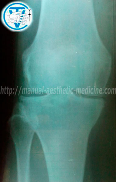
The osteoarthritis of a knee joint is characterized by degenerative lesions of hyaline cartilage.
The cartilage that lies between the contact surfaces of the tibia and the femur becomes thinner, stratified
and eventually loses its cushioning properties. In advanced cases, the disease leads to the complete
disappearance of cartilage; the articular gap is narrowed and it is leading to much more wearing of the
cartilage. As a result, the articular surfaces of the bones are changed and subsequently the osteophytes
start to grow; they known as "spikes". This leads to the joint strain, appearance of severe pain and
limitation of the joint mobility.
MRI of the knee (the stages of research)
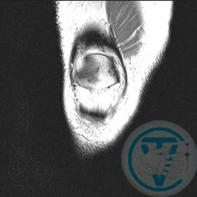
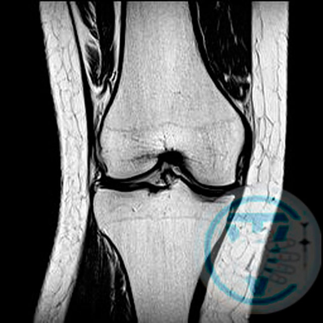
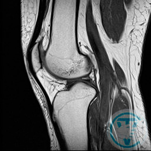
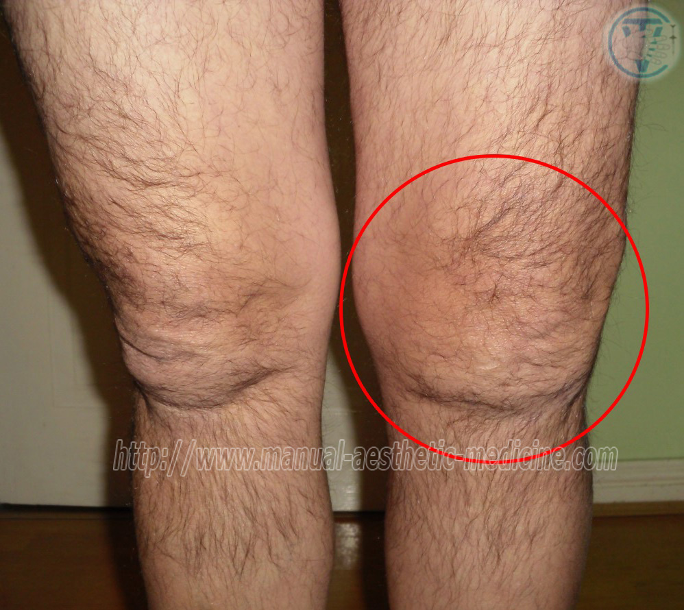
MRI examination of the right knee joint
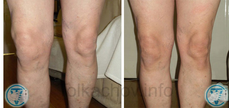
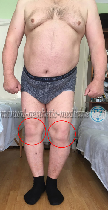
The main symptom of the disease is strong, cutting pain and more often on the inner surface of the joint.
The pain increases with movement or exercise. The disease begins gradually with minor pain or discomfort in the
knee when walking. The pain is worse during the movement, especially during the descent or climbing stairs. Pain
often occurs when the person needs to rise from a seated position; it is difficult to straighten the leg. Later
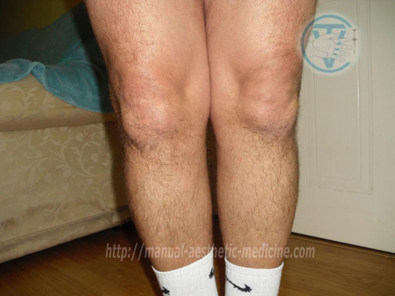
on the deformation, swelling and even lameness manifest in the affected joint. When person is walking the
crunching can be heard by oneself. Hypotrophy of the muscle groups of the femur and tibia is marked not rare.
Contracture and blockade of the joint appear in the later stages of the disease.
Generally, the disease is not limited by one joint only. Later the other joints are involved in the
pathological process such as a hip or knee on the same side usually or other lower limb. Surgical treatment
consists replacing the affected joints by implants. These operations are traumatic and quite often have
different complications. Therefore the development of effective conservative treatment methods is critical.
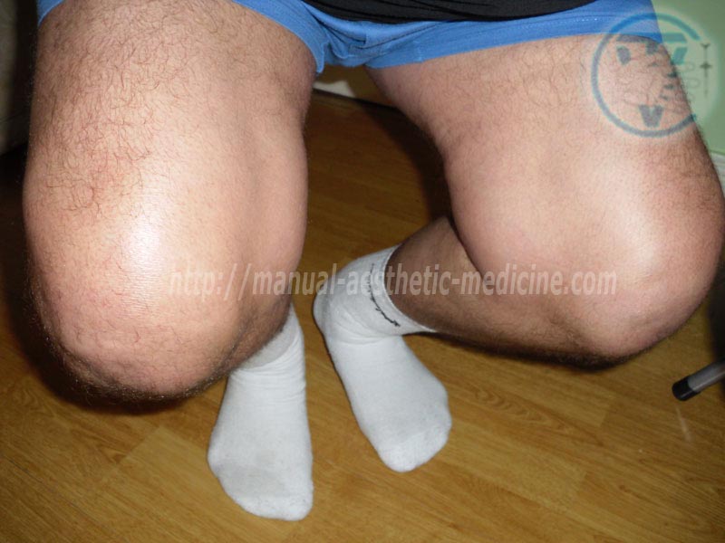
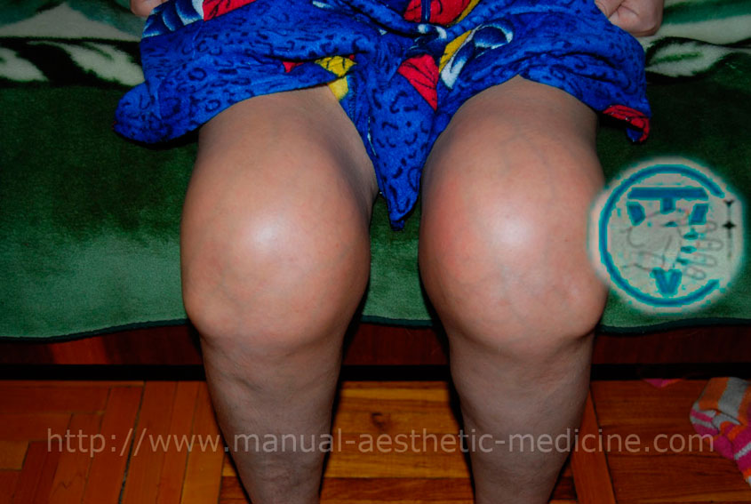
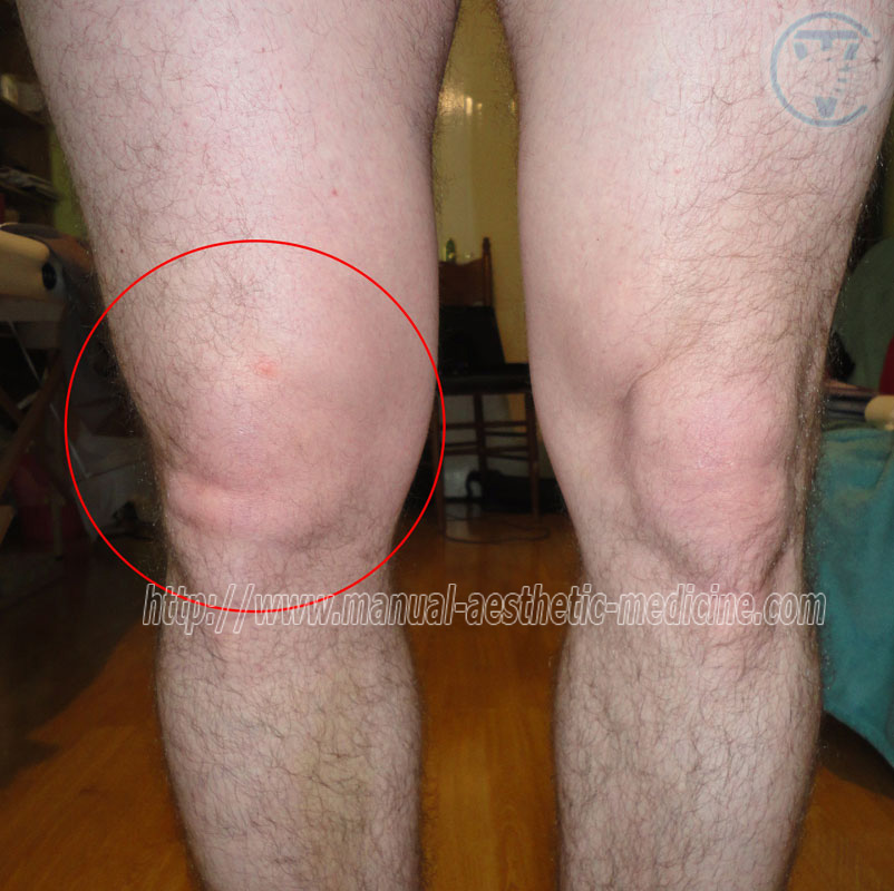
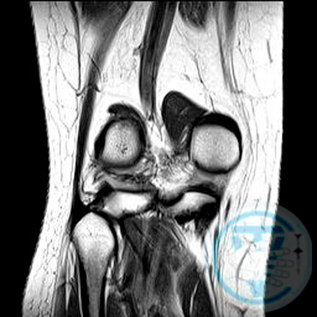
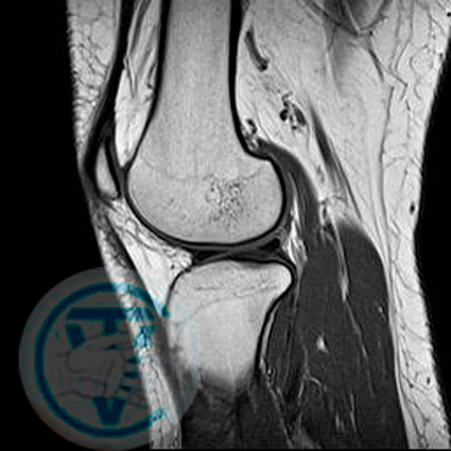
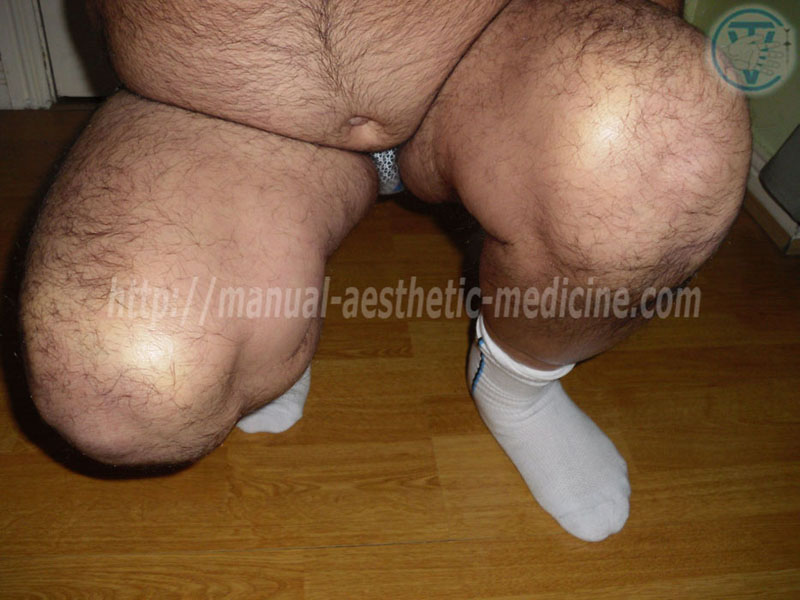
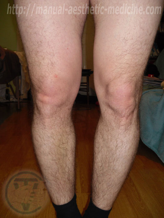
Right knee Osteoarthritis | Arthritis of the right knee joint (bend test)
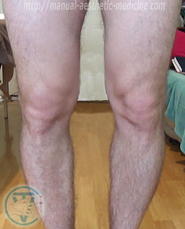
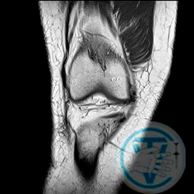
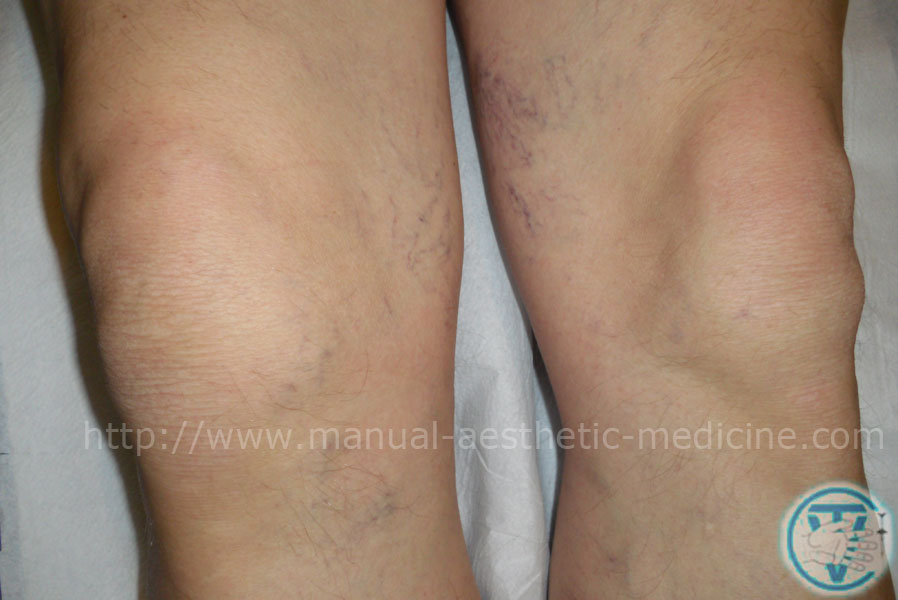
Post-traumatic arthritis of the right knee joint
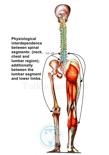
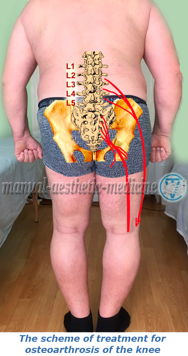
We believe that apart from endocrine and autoimmune processes leading the destruction of the joint an important role belongs to violation of their innervation and blood supply. One of the main reasons causes the breach of innervation can be the infringement of nerve structures (at the L3 – L5 levels of the nerve root exits and also sacral plexus); they are responsible for the neurotrophic regulation of the joint. The treatment is carried out in accordance with “the principles of the multilevel approach in the treatment of chronic and degenerative diseases”, described in our article (medical journal “Bulletin of the science researches”, Ukraine, No. 3 of 2011)". Our treatment is effective; it gives good clinical results with long-term remission and often allows avoiding surgical treatment. The treatment should begin at the early stages of the disease.
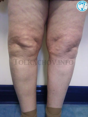
Coxarthrosis
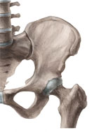
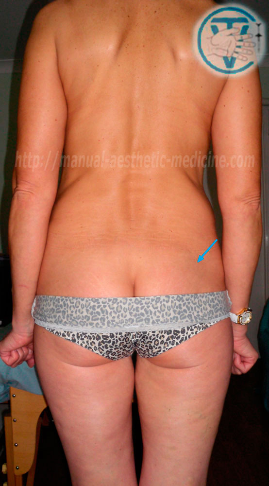
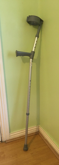
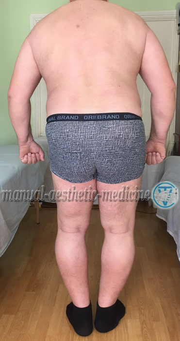
One of the most complicated diseases of the musculoskeletal system is deforming coxarthrosis. The disease of a coxofemoral joint is characterized by destruction of articular surfaces of the femur head and acetabulum, that is followed by distortion of the motor and support functions of lower limbs, increasing painful sensations initially at the beginning of movements and then in the state of rest. Damage of coxofemoral joint quite often leads to distortion of pelvis. Hypotrophy and atrophy of muscles in buttock area, in hip and shin muscular group, decrease of muscular strength becomes noticeable. Further, limping appears. When walking patients are compelled to use a cane or crutches. As a consequence of movement restriction volume in a coxofemoral joint, compensatory increase of mobility in lumbar backbone area becomes apparent. (Treatment is carried out according to "The principles of multilevel approach to treatment of chronic and degenerate disease of a man").
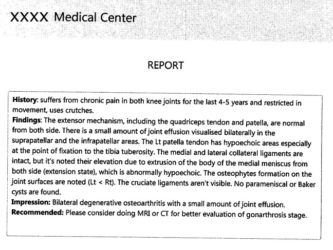
Radiology Result
Patient Xxxxxxx Age / / 1959
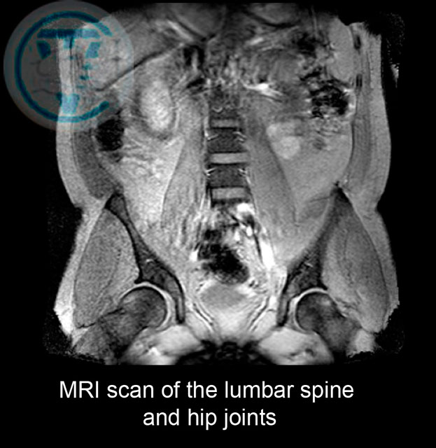
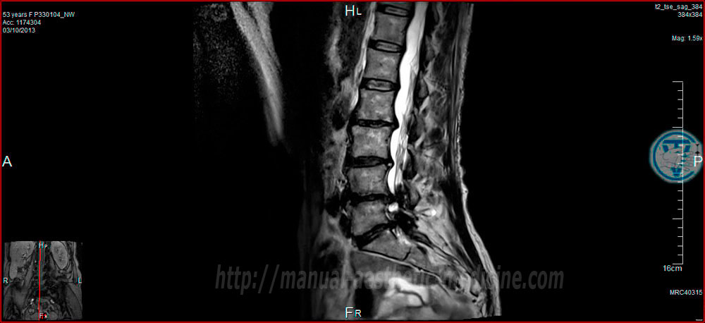
MRI Lumbar Spine / / 2018
Report Details
T1, T2- weigted and STIP sequences were performed in sagital sections in addition to axial T2- weighted images
Normal vertebral alignment.
Moderate degenerative changes with narrowing of the intervertebral disc and osteophytes seen at L5-S1 Level and mild degenerative changes noted in relation to the other
levels of lumbar spine.
1/Osteoarthritis seen in the facet joints of L3-L4, L4-L5 and L5-S1 levels.
No significant focal disc prolapse or spinal canal stenosis nated.
2/ At L4-L5 and L5-S1 levels there is however a mild posterior disc bulge and the combination of this disc with the osteophytes formation and the osteoarthritis in the
facet joints at those two levels caused moderate encroachment on the exit foramina blaterally at those two levels and with no significant spinal canal stenosis.
Narmal appearanca of the conus of the spinal cord.
Radiology Result
Patient Kxxxxx
Age / / 1961
Stadiy Discription MRI SPINE LUMBAR, SACRAL
INDICATION For MRI LUMBAR SPINE ONGOING BACK PAIN. Examination 2019
L1-L2 There is a left pacentral disc bulge which causes mild canal stenosis. No nevral exit foraminal stenosis
L2-L3 There is grade 1 retrolistesis of L2 on L3, There is moderate loss of disc height, disc desiccation , a disc bulge and bilateral fecet joint hypertrophy,
There is mild left nevral exit foraminal stenosis withouth exiting nerv root impingement. No central canal stenosis.
L3-L4 There is moderate loss of disc height, disc desiccation, a disc bulge and bilateral facet joints hypertrophy. There is moderate ctntral canal stenosis, moderate bilateral
lateral recess stenosis and mild narrowing of the right nevral exit foramen, predominantly secondary to facet loint hypertrophy. No exiting nerv root impigement.
L4-L5 There is grade 1 retrolisthesis of disk. There is severe loss of disc height, disc desiccation and endplate irregularity. There is a right foraminal disc protrusion
which causes mild narrowing of the right nevral exit foramen. There is mild left nevral exit foraminal stenosis, secondary to ligamentum flavum hypertrophy. No central canal
stenosis or exiting nerv root impigement.
L5-S1 There is grade 1 retrolistesis of L5 on S1. There is moderate loss of disc height, disc drsiccation, a disc bulge and ligamentum flavum hypertrophy.
There is mild central canal stenosis. Bilateral lateral recess stenosis, mild left nevral exit foramen stenosis and moderate right foraminal stenosis.
No exiting nerv root impingement.
CONCLUSION
Multilevel lumbar degenerative disc disease, most marcet at L4-L5 with mild bilateral foraminal stenosis andat L5-S1 with bilateral lateral recess stenosis and moderate right
foraminal stenosis. No exiting nerv root impingement.

Radiology Result
Patient Name Xxxxxxx
Age / / 1959
Height 186 cm, Weight 150 kg
Report Details
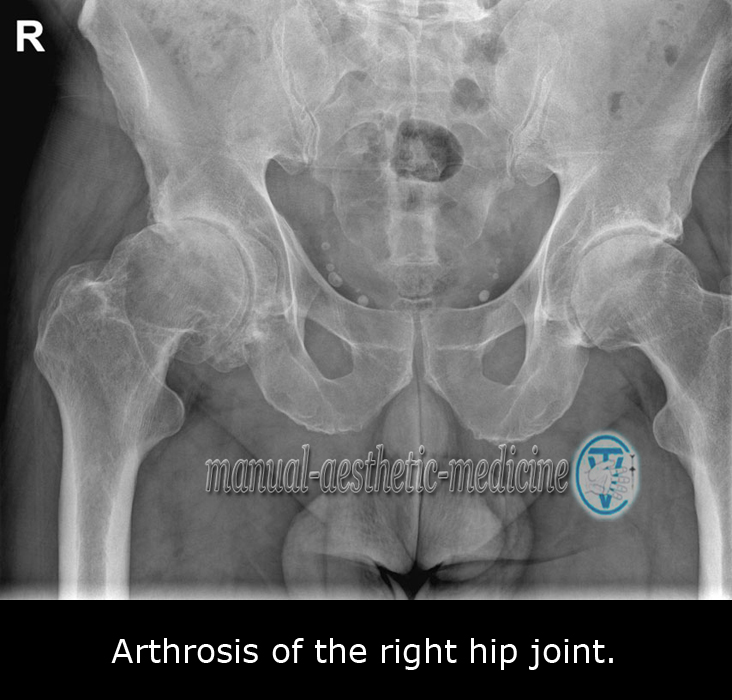
XR PELVIS
Date of Observation 08/ /2018
Results:
XR PELVES
Examination Performed XR PELVIS
Exam Complietion on Date / / 2018
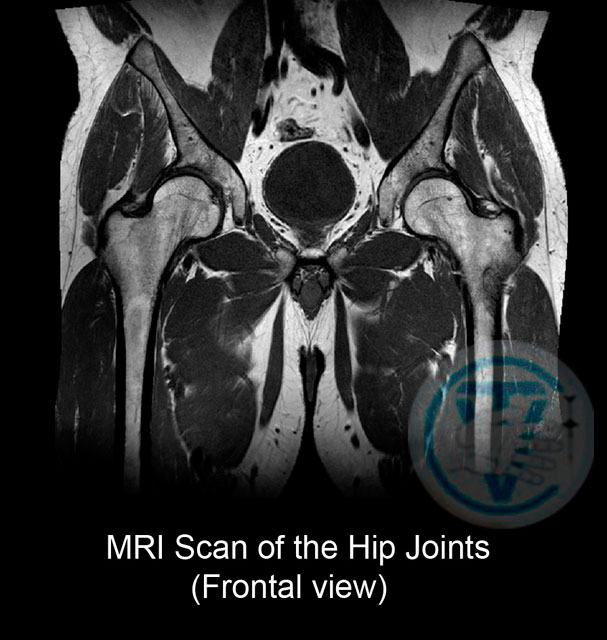
1/ Right Hip marked joint space narrowing and minor deformity
of the 2/ Right femoral head
3/ Marginal osteophyte limping
4/ Subchondral sclerosis
5/ Right hip joint osteoarthritis grade 3
Should be obvious at that stage whether needs surgery, Reiterated advice are weight loss alfuzosin 10 mg 3/12 trial
prostatitis
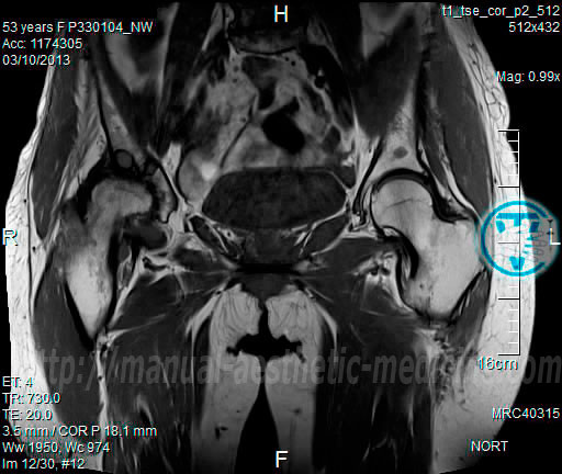
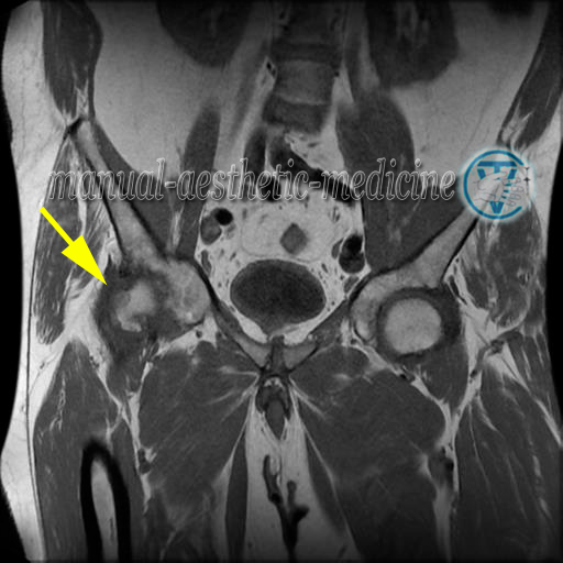
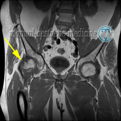
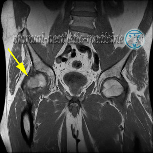
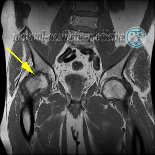
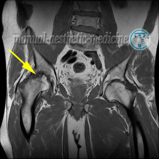
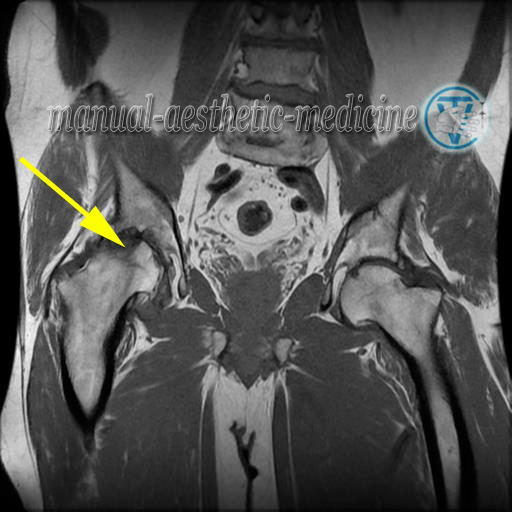
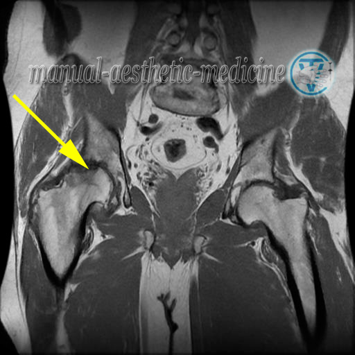
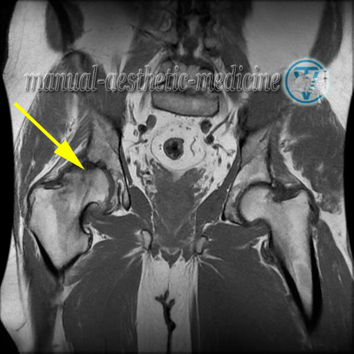
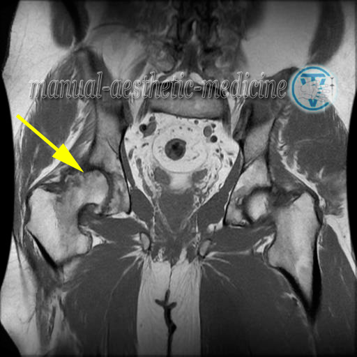
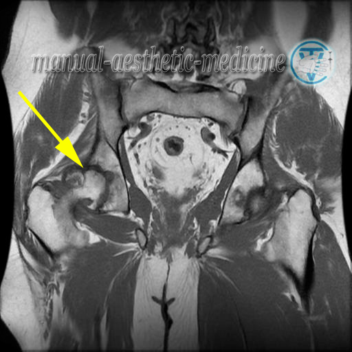
MRI Bony Pelvis / /10/ 2020.
There is marked degenerative change involves the right hip with subchondral cyst formation almost complete loss of joint space and right hip bursitis and small joint effusion. The left hip shows minor degeneration only with no evidence of fracture destructive bone lesion or AVN.
Normal SI joints. The conjoned tendon origins are lagely intact with minor tendinopathy noted on the left.
There is no osteitis pubis.
There is no destructive bone lesion although there is significant liattening of the right femoral head and minor lateral subluxation of the right femoral head noted.
No obvios soft tissue mass.
Concludion Severe drgrnerative change of the right hip noted.
Ultrasound examination 2020
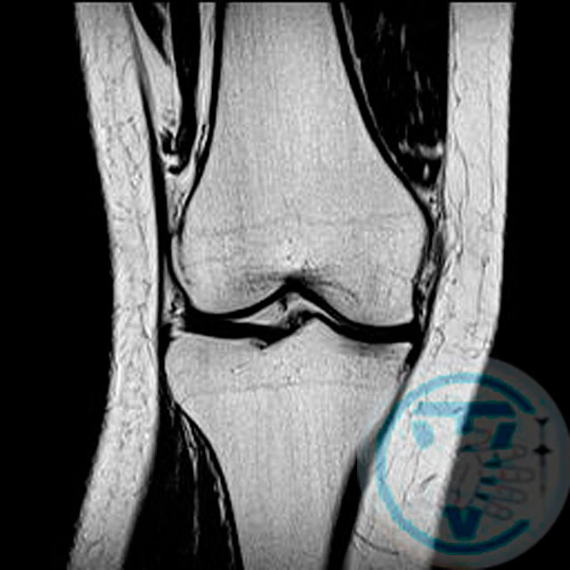
MRI Knee (Right) / /10/2020 MRI of right knee. There is no fracture or destructive bone lesion. A small joint effusion. There is degenerative change at patellofemoral joint space and at the medial joint. Largenperiarticular osteophites are present medially and there is extrusion of medial meniscal tissue with marked degeneration. And likely tear of the medial meniscus. Significant loss of cartilage thickness of the medial joint noted. The lateral meniscus is relatively intact. No soft tissue mass identitied.
References and sources:
http://manual-aesthetic-medicine.com/tpomaitocadd.htmlAdditional pages:
Intervertebral herniation (Hernia of intervertebral disk) Arthrosis deformans of the hip and knee joints (Gonarthrosis) CoxarthrosisSciatica Re-Alignment and Reduction (Vertebral-fat pad, Buffalo Hump) Neck Osteochondrosis (Cervical spondylosis) Carpal tunnel syndrome (Is there a need for an operation?) Visceral Manual Therapy (Key method) SELF-MASSAGE ORGANS OF THE CHEST CAVITY DEFORMATIONS OF FOOT AND TOES
The following are problems my treatment can help with:
- Headaches
- Pain in all parts of the back
- Weak Limbs
- Joints Pain and Stiffness
- Prevention and treatment of problems caused by disc hernia
- Sport Injuries
- “Frozen Shoulder”
- Cellulite and Localised Fat reduction
- Correction of Figure
- Treatment of acne on the face and back (the original technique)
- Asthma
- Chronic Constipation (Treatment)
Manipulation is also possible on the elderly and on pregnant women.
Dr. Vladimir Tolkachov. A man with anaesthetic in his hands...Marty Mc Cool